AV Malformation (AVM) Embolization
Advanced Minimally Invasive Treatment for Arteriovenous Malformations
AV Malformation (AVM) embolization is a highly specialized endovascular procedure used to treat abnormal connections between arteries and veins in the brain or spinal cord. By blocking the abnormal blood vessels, embolization helps reduce the risk of bleeding, alleviate symptoms, and improve the safety and effectiveness of subsequent surgery or radiosurgery when required.
Our expert neurointerventional specialists provide advanced AVM embolization using state-of-the-art imaging technology and minimally invasive techniques to achieve the best possible outcomes.
What is an Arteriovenous Malformation (AVM)?
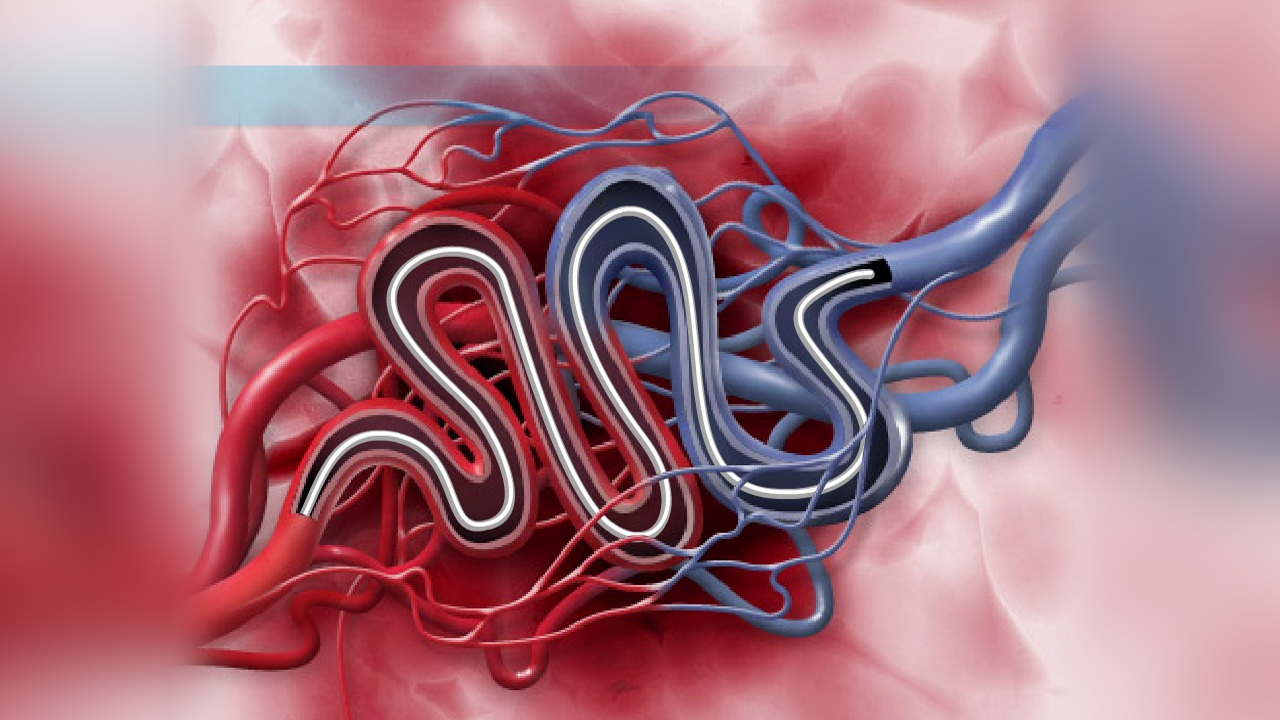
An arteriovenous malformation (AVM) is a congenital vascular abnormality in which arteries connect directly to veins without the normal network of capillaries. This abnormal connection creates high-pressure blood flow that can weaken blood vessels and increase the risk of rupture and bleeding.
AVMs can occur in the brain, spinal cord, or other parts of the body, but cerebral AVMs are among the most serious due to their potential to cause stroke or brain hemorrhage.
Why is AVM Embolization Performed?
AVM embolization is performed to reduce blood flow through the malformation and decrease the risk of complications. The procedure may be used to:
- Reduce the risk of brain hemorrhage
- Treat symptomatic AVMs
- Decrease AVM size before surgery
- Improve the effectiveness of radiosurgery
- Reduce neurological symptoms
- Minimize blood loss during surgical removal
- Provide definitive treatment in selected cases
Symptoms of an AVM
Some AVMs may remain asymptomatic for years, while others may cause:
- Severe headaches
- Seizures
- Weakness or numbness
- Vision problems
- Difficulty speaking
- Dizziness
- Balance and coordination issues
- Memory problems
- Brain hemorrhage
- Stroke-like symptoms
How is AVM Embolization Performed?
- Step 1: Comprehensive Evaluation
- MRI Brain
- CT Scan
- CT Angiography (CTA)
- MR Angiography (MRA)
- Digital Subtraction Angiography (DSA)
- Step 2: Catheter-Based Access
A microcatheter is inserted through an artery in the wrist or groin and navigated to the arteries feeding the AVM. - Step 3: Embolization
Specialized embolic agents such as liquid embolics, particles, or coils are delivered into the abnormal blood vessels to block blood flow to the AVM. - Step 4: Post-Procedure Assessment
Imaging is performed to confirm successful reduction or occlusion of the AVM and evaluate treatment effectiveness.
Diagnostic studies may include:
Benefits of AVM Embolization
- Minimally invasive procedure
- Reduced risk of hemorrhage
- Improved neurological outcomes
- Reduced AVM size
- Enhanced surgical safety
- Better radiosurgery planning
- Shorter recovery time
- Reduced hospital stay
- Preservation of surrounding healthy brain tissue
Recovery After AVM Embolization
Most patients recover within a few days, depending on the complexity of the procedure.
Recovery typically includes:
- Neurological observation
- Follow-up imaging studies
- Medication management
- Activity modifications
- Scheduled specialist consultations
Risks and Potential Complications
Although AVM embolization is generally safe, potential risks may include:
- Stroke
- Bleeding
- Blood vessel injury
- Brain swelling
- Temporary neurological symptoms
- Infection
- Allergic reaction to contrast material
Why Choose Our Neurovascular Center?
- Expertise in AVM diagnosis and treatment
- Advanced endovascular embolization techniques
- State-of-the-art neuroangiography facilities
- Comprehensive cerebrovascular care
- Personalized treatment planning
- Multidisciplinary team of neurointerventionalists, neurosurgeons, and neurologists
- Advanced stroke and neurovascular services
We are dedicated to providing innovative and effective treatment solutions for patients with complex vascular disorders of the brain and spinal cord.